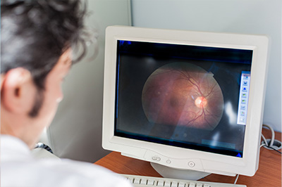
Annual wellness screening is performed for all adult patients so the doctor can obtain a baseline and screen for many pathologies. It consists of 3 simple procedures ( retinal camera, optical coherence tomography (OCT) & visual field testing) as they may not be covered by your insurance plan if your eyes are healthy.
At North Country Optical, we maintain a position at the forefront of advances in care to help every patient protect and preserve optimal eye health and vision. We firmly believe an Annual Eye Wellness Screening offers one of the best ways to monitor your eye health, diagnose issues early in their onset, and prevent vision loss.
Why get Eye Wellness Screening every year?
Taking small steps to protect your eye health and vision goes a long way. An Annual Eye Wellness Exam offers the valuable information required to diagnose problems early in their onset when management and care can most effectively treat, resolve, halt, or significantly slow progression to prevent vision loss and even blindness.
Remember, many eye diseases, like macular degeneration, diabetic retinopathy, glaucoma, and more, do not exhibit overt symptoms in their early stages. Plus, the back of your eye provides important insights into your overall health. Evidence of conditions like high blood pressure, high cholesterol, diabetes, can also be detected.
A small investment in the long-term health of your eyes
Today, thanks to advances in optical technology, it’s possible to get insights into your eye health and vision with the ultimate precision.
While eye wellness screening is not typically covered by insurance, this affordable procedure represents a small yet highly important investment in enjoying the benefits of optimal eyesight.
Optical Coherence Tomography
Optical Coherence Tomography (OCT) provides a high-resolution and cross-sectional view of the back of the eye and retina.
It provides detailed images helpful in detecting retinal conditions, optic nerve disorders, and visualizing the eye’s anterior segment, including the cornea, anterior chamber, iris, and angle.
Retinal Photography
Retinal photography captures a broad digital view of the back of the eye. It provides high-resolution and high-definition images of the retina, optic nerve, small blood vessels of the eye, and other features to permit an accurate assessment of eye health as well as the diagnosis of various ocular, neurologic and systemic disorders. It also facilitates documenting the progression and response to treatment of conditions such as age-related macular degeneration, diabetic retinopathy, glaucoma, ocular disorders, retinal neoplasms, and choroid disturbances. Best of all, we can do all this without having to place eyedrops to dilate your eyes.

